

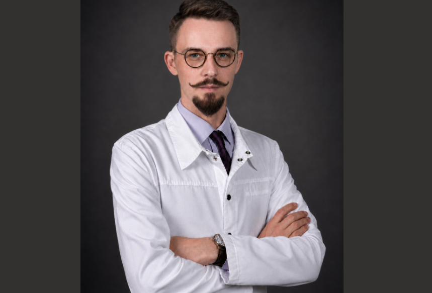
Dr. Michael Lebenstein-Gumovski, PhD is Senior Scientific Officer and Neurosurgeon, in the Neurosurgery Department, of the Sklifosovsky Clinical and Research Institute for Emergency Medicine, Moscow, Russian Federation (https://sklif.mos.ru/), where his team is engaged in both neurosurgical and experimental practice, conducting advanced research in the field of spinal cord injury restoration, spinal cord transplantation and head transplantation.
Since 2013, Dr. Lebenstein-Gumovski has been studying spinal cord injury, and also developing methods for restoring the full functional and morphological repair of the spinal cord.
Dr. Lebenstein-Gumovski’s work is aimed at studying the effect of fusogens on nervous tissue, developing new methods and techniques for treating spinal cord injury, developing methods for its resection and transplantation. The lab develops and studies various methods of neuroprotection, combining methods to achieve better results and the current focus is the study of combination fusogen-induced (PEG-chitosan, Neuro-PEG) axonal restoration of the spinal cord after its complete transection.
SSN: What drove you to choose a career in spinal research?
M L-G: Neurosurgery has always attracted me because of its breadth and complexity. We work with blood vessels, tumors, bone, and infections. We work with the organ that, in many respects, defines us as human beings—the brain—and with the structures that govern the body: the spinal cord and peripheral nerves. That combination is endlessly fascinating.
As for the specific problem my team and I study, neurosurgery still contains several central challenges that are often described as “unsolvable.” Spinal cord injury is one of them. In my view, this is not because neurosurgeons lack technical skill, but because surgical practice has too often been separated from neurobiology. For many years, restoration of neural tissue was considered impossible, until the mechanisms of axonal growth and neuroplastic remodelling were identified and began to be understood. I kept asking myself why, despite the existence of regenerative mechanisms, the spinal cord remained so resistant to restoration. The answer “it is impossible” never satisfied me. I felt that a different perspective, and a fundamentally different approach, were needed. Hundreds of thousands of spinal column and spinal cord injuries occur worldwide every year, and behind that statistic are individual human tragedies.
At the same time, as a student, I read the work of Robert White and Vladimir Demikhov on head transplantation, where the central obstacle was also the inability to restore a transected spinal cord. My compatriot, the science-fiction writer Alexander Belyaev, wrote a novel about head transplantation more than a century ago; in our country, it was familiar to almost every schoolchild. Of course, I was not pursuing head transplantation as a practical objective at that time. But I did understand that solving the spinal cord problem could, in principle, transform even the most radical questions in neurosurgery.
A certain degree of ambition, a healthy scientific curiosity, and the desire to approach fundamental biological mysteries have always been among the main engines of my scientific work.
SSN: Along with a team of international researchers, you have helped restore motor, sensory and pelvic functions after complete spinal cord transection in animal models using fusogenic therapy. Could you tell us more about your research and findings?
M L-G: Over the past two decades, many studies of cellular technologies have produced results that are important but, in my view, not sufficiently transformative for severe spinal cord injury. When I first began studying this field, the work of Borgens, Cho, and Bittner attracted my attention because it offered a genuinely different principle: membrane fusion in injured peripheral nerves. I remember thinking: this may be the key. Why attempt only to grow new axons if we can also try to fuse the damaged ones?
In 2013, I attempted to adapt Bittner’s protocols to spinal cord injury. The initial results, however, were neither satisfactory nor reproducible. That failure was important, because it forced us to experiment more actively. Around that time, a team began to form around the project, including chemists, pathologists, and my clinical colleagues. Together, we went through many experimental difficulties before obtaining results that could be discussed with confidence.
We first developed several combinations of different substances, modified Bittner’s protocols, and identified key conditions required for applying this approach to the spinal cord rather than to a peripheral nerve. Eventually, this work led to a broader technological platform that we now describe as fusogenic neurosurgery.
The principle can be explained quite simply. When a nerve is injured, axons that originate from neurons in the spinal cord are interrupted. Imagine a tree branch cut in the middle. One part of the branch dies, while the other remains connected to the tree and can continue to grow. A similar process occurs in the nervous system: the distal axonal segment undergoes degeneration, while the proximal segment becomes a stump. If two segments of a peripheral nerve are approximated and sutured, axons can regenerate at approximately 1–4 mm per day and eventually begin transmitting impulses again from the neuronal cell body to the muscle. This regeneration is supported, among other factors, by the glial environment surrounding the axons.
In the spinal cord, however, this process does not occur in the same way. A cascade of destructive changes rapidly develops in neural tissue. Secondary injury and edema appear, and immune cells and immune complexes enter the damaged area. In practice, the lesion—whether it results from transection or from crush injury, as is often seen clinically—can expand severalfold within a few hours. Within weeks, the biological debris may be replaced by a cyst surrounded by a glial scar and connective tissue. This creates both a physical barrier to axonal growth and a biological environment in which the distal axonal segments have already degenerated and the neural tissue has undergone profound remodelling.
In our experimental model, we transect the spinal cord and, without allowing these degenerative processes to develop, immediately cool the tissue, irrigate it with a sequence of specialized solutions, and apply the fusogen. The fusogen is central: it induces the membranes of transected axons to fuse again. This is not a one-to-one reconnection—that would be impossible. Rather, fusion appears to occur within functional groups: motor fibers with motor fibers, sensory fibers with sensory fibers. Returning to the tree analogy, imagine that after cutting a branch, we prevent the cut surfaces from dying, refresh them quickly, bring them into precise and tight apposition, and seal the junction with a special adhesive. After some time, the branch again receives nourishment and continues to live.
Colleagues have often said to me, “But our patients do not have a cleanly transected spinal cord; they have a crushed spinal cord.” That is true. Precisely for this reason, we believe that a destroyed segment filled with biological debris, damaged cells, and nonviable axons may need to be removed, and that fusogenic technology should be applied to healthy, freshly prepared spinal cord ends. This implies a serious surgical strategy, but it may be the type of strategy required for meaningful recovery. Because fusogens require two viable axonal ends that can be brought into apposition and fused, chronic and long-standing injuries are not suitable for this form of therapy: by then, the relevant axons have already degenerated.
It is important to emphasize that fusogens are not science fiction. Membrane fusion has been studied for more than 50 years. In cell biology, polyethylene glycol is routinely used to induce the fusion of mononuclear cells into multinucleated cells. Similar biophysical mechanisms operate in the axons of certain fish, amphibians, and roundworms, allowing them to repair injuries to the nervous system. In those organisms, this process is mediated by protein fusogens. In mammals, as with many ancient biological capacities, this mechanism appears to have diminished during evolution. Our goal is to reactivate, or at least imitate, that capacity through external intervention.
We began with rat models and obtained reproducible effects, although I would not describe those results as highly encouraging. We then applied the technology in rabbits, expanded the protocol, and obtained a remarkable result: within a month, the rabbits were jumping again. After that, we moved to the most translational model available to us: pigs. The porcine spine and spinal cord are close to the human system in neuroanatomy and biomechanics. In our first pilot study in 2022–2023, in which we applied the most complete version of the protocol, pigs with complete spinal cord transection began moving their limbs within the first weeks.
We have been extremely fortunate with our team. Our chemists approach the biosynthesis of new fusogen variants creatively; our surgical team is willing to develop new operative ideas, refine procedures, and provide meticulous postoperative care for the animals. The fact that we can see, with our own eyes, taboos beginning to collapse gives us tremendous motivation to continue.
I should also stress that no fusogen, however powerful, will work in isolation. Even if a colleague were to take a vial from my laboratory and attempt to use it, nothing meaningful would happen. The essential element is not the fusogen alone, but a strict surgical algorithm and a combination of technologies and methods that sequentially prepare the spinal cord for fusion and continue to influence it after surgery. This system is the most valuable part of our technology. If the nuances are not followed, the fusogen is useless. It is rather like bringing a quantum computer into a forest hut: it may be extraordinary, but in that environment it simply stands there unused.
SSN: How could your findings help reshape spinal cord function and the patient experience?
M L-G: If we succeed in bringing this technology into clinical practice, we will unfortunately not be able to cure the millions of people already living with chronic spinal cord injury. As I noted earlier, membrane fusion requires viable axonal ends in close apposition, and those conditions are no longer present several days after injury. The more realistic objective is therefore to prevent newly injured patients from developing profound, permanent disability by applying the technology within the first hours after spinal cord injury.
To make that possible, existing protocols for the management of these patients would have to be substantially revised, including referral pathways, prehospital criteria, diagnostic algorithms, and surgical logistics. Time is critical. The longer it takes to bring a patient to the operating room, the fewer axons are likely to remain available for restoration.
For an initial clinical effect, it may be sufficient to restore only 10–15% of functionally significant white-matter fibers in the spinal cord. After that, gray matter networks and interneuronal circuits may participate by redirecting part of the signal and functioning, in a sense, as biological relays. This interneuronal mechanism is one form of neuroplastic remodeling that can improve functional outcomes over time. Ultimately, neuroplasticity and rehabilitation will play decisive roles in recovery. We should not promise complete restoration of function. But if patients are no longer confined to a wheelchair and can regain a greater degree of independence and social participation, that would already represent a profound change in their lives.
SSN: Are you currently involved in any other research projects? If so, could you tell us more about its focus?
M L-G: At present, most of my time, together with the team, is devoted to fusogenic research. One of our current projects is designed to answer a very practical question: which fusogen and which protocol are the most effective? There are many fusogens, and different groups have studied different concentrations, molecular weights, exposure times, and even temperatures. Most of these studies have been conducted in peripheral nerves. Yet no unified protocol exists. We want to provide a rigorous comparative answer.
We are therefore conducting a large screening study divided into two preclinical phases. In the first phase, we will select the strongest candidates from approximately 40 different protocols. In the second phase, we will test those protocols in a large sample of translational models—pigs—using the same technological principles that would be required in human application. If the results are sufficiently convincing, we are prepared to begin clinical studies in patients within our hospital setting.
As invited experts, we are also involved in research on the transplantation of olfactory ensheathing cells in chronic spinal cord injury.
In parallel, we are participating in clinical studies evaluating the neuroprotective effects of umbilical cord blood-derived mesenchymal cells in limiting secondary injury after spinal cord trauma. These directions are overseen by Professor A. Grin, Chief Neurosurgeon of Moscow and Head of the Department of Neurosurgery at the Sklifosovsky Institute. Naturally, we also have several other projects, including studies related to neural tissue transplantation, that I cannot disclose at this stage.
In addition, I serve as the lead scientific consultant for the Dowell Bio project, which investigates fusogenic technologies at other research sites and reproduces our protocols. This work may substantially accelerate global research in the field. We are also developing brain perfusion systems, although that direction remains at a very early stage.
SSN: How does the future look in treating spinal cord injury?
M L-G: In Russian, we say, “The ice has broken.” In this field, we have not simply pushed the wall of taboo a little farther away; we have begun to dismantle it using the tools of evidence-based science. I am confident that within the next 10–15 years, at the intersection of fusogenic therapy, cell-based technologies, gene therapy, and electrical stimulation, we will find a convincing answer to spinal cord injury, both acute and chronic.
It is more difficult to predict how quickly these advances will enter everyday clinical practice. A number of legal, regulatory, and organizational barriers will have to be addressed before such technologies become widely available. But I am less concerned about legal barriers than about the persistence of stereotypes among some practicing surgeons regarding what is supposedly “impossible.” Those intellectual barriers can be even harder to overcome.
SSN: What do you enjoy most about your work?
M L-G: My work combines the roles of scientist, surgeon, and teacher. As a scientist, I enjoy the elegant interplay of cells, molecules, and tissues. Sometimes my team and I are as excited as children when we see a beautiful neuron under the microscope in a place where, after injury, it should no longer exist.
In neurosurgical practice, what I value most is the operation itself—the moment when one approaches something close to the act of creation. It is a moment of truth. And in teaching, I enjoy seeing talented students sometimes surpass their teachers. There is nothing wrong with that. It is natural, and it is how progress occurs.
SSN: … and the worst?
M L-G: Death and ruined lives. Unfortunately, there is still too much of both. No matter how long one works in this field, it is impossible to become reconciled to that. I still have to tell patients that they will remain disabled forever and will never again move their legs or arms. We must have courage, resilience, and composure, but that does not make it easy.
SSN: What has been the highlight of your career so far?
M L-G: The most important thing is that the work of our team is beginning to bear fruit, and that what was once considered impossible is becoming achievable. It is probably not a single moment, but rather a process that has begun recently through the coordinated efforts of many people, both within and beyond our own group.
SSN: Are you planning to attend or speak at any medical conferences or events over the next year?
M L-G: I have not yet made specific plans. I may attend the EANS Congress if I am not occupied with experiments. I regularly attend local forums and, when time allows, occasionally give lectures at universities. If there is sufficient time and interest, perhaps we will prepare something more substantial. After all, we now have something important to discuss.
SSN: If you didn’t work in the health industry, what would you be?
M L-G: It is difficult for me to imagine myself doing anything else. From early childhood, I knew with certainty that I would work in medicine. At the same time, I have a deep interest in art, especially Italian painting, and I occasionally practice painting myself. It brings harmony to life, helps me look at certain things from unexpected angles, and trains fine motor skills, which are essential in surgery.
SSN: What advice would you tell your 21-year-old self?
M L-G: Keep going in the same direction, but be bolder.
SSN: How do you think the future looks within the field of spinal surgery and what are your predictions for 2027 and the next decade?
M L-G: I think spine surgery will continue to move toward minimally invasive approaches, and implants will become more biocompatible, biodegradable, and biointegrative. But the fundamental concepts of spine surgery are unlikely to change radically, because the spine is, above all, a structure of mechanical function.
The spinal cord is different. In the next five to ten years, I believe important discoveries will be made, and key fundamental principles will be confirmed, that will substantially change our understanding of what spinal cord recovery can mean. Artificial intelligence and new methods in neuroscience are now producing a global leap forward, and that cannot fail to influence this field.
My teacher and mentor, Professor Grin, has always said: “With the spine, we have learned to do almost everything—shorten it, fix it, and replace parts of it with implants. With the spinal cord, we still cannot do anything. The future of neurosurgery lies where that boundary is crossed.”
In another 10–15 years, I believe the chronicle of a new spinal cord surgery will have begun to take shape. For now, we are still writing its alphabet.
https://www.researchgate.net/profile/Michael-Lebenstein-Gumovski
