
An international team of researchers has restored motor, sensory, and pelvic functions after complete spinal cord transection in animal models. To achieve this, the researchers used a combined protocol consisting of fusogenic therapy and comprehensive rehabilitation. The study results were published in the peer-reviewed journal PLOS One.
The need for a new therapy
Following severe spinal cord injury, the axons of mammals, including humans, inevitably degenerate, edema develops, neurons die, and currently available methods aimed at preventing these processes remain ineffective. Consequently, spinal cord injury currently represents a critical challenge in neurosurgery. In recent decades, there has been growing interest in a new class of substances – fusogenic sealants – that can induce axonal membrane fusion, restoring their integrity. This energetically efficient repair mechanism is observed in amphibians through the action of fusogenic proteins and can be artificially initiated in higher animals using synthetic compounds. Polyethylene glycol, capable of inducing membrane fusion, and chitosan, which serves as a biomatrix and a neuroprotective agent, form the basis of this method.
Background of the study
A team of scientists led by neurosurgeon Michael Lebenstein-Gumovski, Senior Scientific Officer at the Sklifosovsky Research and Clinical Institute for Emergency Medicine, MD, PhD, has been studying the effectiveness of fusogen-based membrane fusion technologies for over 10 years. In addition to synthesizing a fusogenic compound — a polyethylene glycol-chitosan conjugate (Neuro-PEG) — the researchers have developed a comprehensive technology for fusogenic repair and multi-component rehabilitation for spinal cord injury, which is inextricably linked to successful recovery. The mechanism of action of fusogens is not based on regeneration through axon growth or the generation of new neurons, but solely through the primary fusion of axonal membranes, which is partially stochastic in nature and cannot yet be controlled with absolute precision.
The study’s unique features
A new study titled “Fusogen-induced recovery of spinal cord function and morphology after complete transection” examined the effects of fusogenic therapy on the functional and morphological recovery of the spinal cord following complete transection at the thoracic region. Hungarian Mangalica pigs underwent complete transverse transection of the spinal cord at the thoracic level, followed by spinal fixation using a titanium construct. In the experimental group, the fusogen compound Neuro-PEG was applied directly to the injury site, while another fusogen was administered intravenously; the other group served as a control without treatment. The postoperative period lasted 60 days and included a multi-component rehabilitation program.
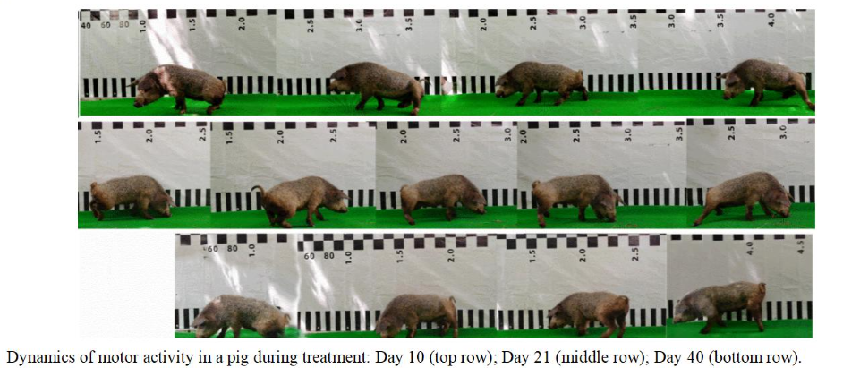
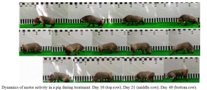
Following fusogenic treatment, sensitivity in the limbs began to return as early as the second day after surgery; by the fifth day, the animals had regained control over urination; by the second week, all animals in the treatment group demonstrated active hind limb movements and attempts to stand up. By the end of the study, the treated animals were able to move independently on all four limbs, although gait instability persisted, whereas control animals showed no movement or sensitivity in their limbs throughout the experiment and exhibited impaired pelvic function.
The results were analysed using standard neurological scales, and statistically significant differences were found between the groups. Tissue preparations revealed a high number of axons crossing the injury site in the experimental group, while degenerative post-traumatic changes were observed in the control samples.
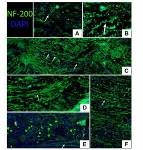
Immunofluorescence microscopy of the spinal cord in the experimental group. NF-200-positive structures are shown in green and nuclei are stained with blue DAPI. The arrows indicate axonal structures crossing the injured area.
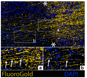
In vivo tracing of spinal cord axons in an animal from the treatment group. The ends of the injury site are marked with asterisks. Nuclear staining with DAPI (blue); FluoroGold (yellow) tracing reveals axons. Arrows indicate axons crossing the injury site in fields a and b (×400).
Experimental results, limitations, and prospects for further research
Analysis of the study results revealed a significant therapeutic effect of fusogenic therapy and the rehabilitation procedures used. The researchers succeeded in restoring physical mobility in the experimental animals compared to the untreated group, whose condition showed no improvement by the end of the 60-day experiment. Furthermore, the scientists hypothesized that optimal fusogenic action and successful axon fusion also depend on the precision of the surgical transection, neuroprotective strategies such as local hypothermia, and the impact on the cellular microenvironment.
However, the authors emphasize that this is an early preclinical study with a small sample size, and further research will be needed to confirm the reproducibility of the results, clarify the underlying mechanisms, and determine the components of the combined protocol required for recovery.
“We are also developing new surgical concepts and have working prototypes of a specialized device designed to precisely transect and align the spinal cord. In the future, if this technique is validated, it should become a comprehensive technological system rather than a single drug or molecule. It will include fusogenic agents, specialized devices, surgical techniques, perioperative neuroprotection, rehabilitation, and patient care pathways,” noted Dr. Michael Lebenstein-Gumovski, the study’s lead author.
It is important to note that the authors do not expect this approach to be applicable to chronic spinal cord injuries, where nerve tissue degeneration and secondary remodeling have already occurred. The concept of fusion is most relevant during the acute phase, when only a few hours have passed since the injury and structural integrity may still be biologically significant. To this end, a spinal cord rehabilitation concept based on debridement theory is being developed in collaboration with Andrey Grin, MD, Doctor of Medical Sciences, Professor, Corresponding Member of the Russian Academy of Sciences and Head of the Neurosurgery Clinic at the Sklifosovsky Research and Clinical Institute for Emergency Medicine.
Research into the fundamental mechanisms of fusogenic restoration of neural tissue is important for the development of neurosurgery and neurobiology and helps humanity move closer to solving the global problem of disability caused by spinal cord injuries. However, it is already clear that the era of fusogenic neurosurgery is beginning.
The authors have also published a preprint of the first-ever comprehensive systematic review with a meta-analysis on fusogens, covering more than 50 years of fusogen use and 26 years of research on their effects on nervous tissue, providing a thorough fundamental overview of their mechanisms of action (Fusogens for Axon Repair in Spinal Cord and Peripheral Nerve Injuries – Studies, Methods, and Mechanisms (systematic review with meta-analysis): https://www.biorxiv.org/content/10.64898/2026.03.20.712959).
Reference: Lebenstein-Gumovski M, Rasueva T, Kovalev D, Canavero S, Zharchenko A, Petrov P, et al. (2026) Fusogen-induced recovery of spinal cord function and morphology after complete transection. PLOS One 21(6): e0349579. Fusogen-induced recovery of spinal cord function and morphology after complete transection | PLOS One
Source: Sklifosovsky Research and Clinical Institute for Emergency Medicine
Images: Photographic materials (used with approval by the authors):
– Video footage demonstrating the recovery of motor activity in treated animals;
– Immunofluorescent images of neurofilament-positive fibers in the area of injury;
– Light microscopy and FluoroGold tracing images.
